Animal Cell Diagram Cytoskeleton / Interactive Eukaryotic Cell Model : The walls, hull, and overall structure of the ship is like the cytoskeleton of a cell because they give the ship its shape just like how the cytoskeleton gives the cell its shape.
byHedy Howlett-0
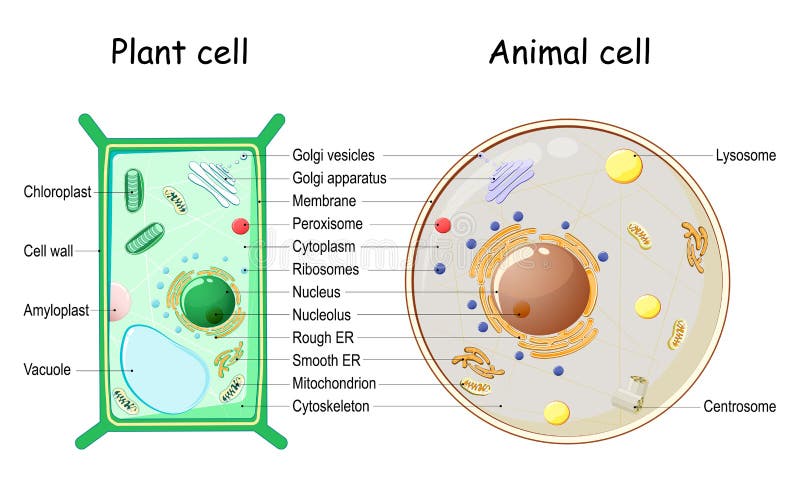
Animal Cell Diagram Cytoskeleton / Interactive Eukaryotic Cell Model : The walls, hull, and overall structure of the ship is like the cytoskeleton of a cell because they give the ship its shape just like how the cytoskeleton gives the cell its shape.. Cell organelles structure and parts. The diagram, like the one above, will include labels of the major parts of an animal cell including the cell membrane, nucleus, ribosomes, mitochondria, vesicles, and cytosol. The walls, hull, and overall structure of the ship is like the cytoskeleton of a cell because they give the ship its shape just like how the cytoskeleton gives the cell its shape. The diagram shows an idealized cell: The cytoskeleton is a network of filaments and tubules found throughout the cytoplasm of the cell.
Cytoskeleton, a system of filaments or fibers that is present in the cytoplasm of eukaryotic cells. Microfilaments are the thinnest of all the cytoskeletal. Animal cells have one or more nucleoli, but some cell types do not have any. A tour of the animal cell by biology professor dr. Microtubules, microfilaments and intermediate filaments.
Plant Cell And Animal Cell Structure Stock Vector Illustration Of Mitochondria Cell 189373848 from thumbs.dreamstime.com Animal cell to cruise ship analogy > . In animal cells the mtoc is called the centrosome. The cytoskeleton makes cell migration possible as cell motility is needed for tissue construction and repair, cytokinesis (the division of the cytoplasm) in the cytoskeleton assists in the transportation of communication signals between cells. Since nucleoli carry out the production and maturation of ribosomes, large numbers of ribosomes are found inside them. An animal cell diagram is a great way to learn and understand the many functions of an animal cell. Thomas risler institut curie, centre de recherche, umr 168. The cell cytoskeleton serves to protect the cell from both pulling (tensile) and pushing (compression) stress , so maintaining the cell tensegrity. Unlike the eukaryotic cells of plants and fungi, animal cells do not have a cell wall.
It gives cell shape, organizes organelles, involves molecule in this figure left:
A cytoskeleton is present in the cytoplasm of all cells, including bacteria, and archaea. The diagram shows an idealized cell: Several cellular structures are built around a core of cytoskeletal proteins. In the complete animal cell centrosome, the two centrioles are arranged such that one is perpendicular to the other. The nuclear lamina forms an organized meshwork on the internal face of the the diagram includes 8 rna polymerases however the number can vary depending on cell type. During animal cell division, the centrioles replicate (make new copies) and the centrosome divides. Animal cell to cruise ship analogy > . Perhaps the best known examples are cilia and flagella. A tour of the animal cell by biology professor dr. The cytoskeleton of a biological cell is the framework of tiny tubes and filaments that forms the internal structure of the see the diagram to see how these are arranged. This function is especially important in animal cells, which lack walls. In reality, actin arrays are interconnected in various. 5 metaphase anaphase telophase kinetochore microtubule early metaphase metaphase.
Summary of the structure and function of a eukaryotic cell. The diagram shows an idealized cell: Perhaps the best known examples are cilia and flagella. The cytoskeleton is closely involved in many processes including cell division, growth cytoskeleton consists of three types of elements: In animal cells, two networks of intermediate filaments provide the nucleus with mechanical support:
1 Animal Cells The Cytoskeleton Licrotubules In Green And The Actin Download Scientific Diagram from www.researchgate.net The walls, hull, and overall structure of the ship is like the cytoskeleton of a cell because they give the ship its shape just like how the cytoskeleton gives the cell its shape. This structure is where microtubules are assembled and anchored. What does the cytoskeleton provide? In the complete animal cell centrosome, the two centrioles are arranged such that one is perpendicular to the other. The cytoskeleton of a biological cell is the framework of tiny tubes and filaments that forms the internal structure of the see the diagram to see how these are arranged. After fixation and labelling with specific probes. It also serves as the attachment point for both the intracellular cytoskeleton and, if present, the cell wall. Among these, vertebrate skeletal muscles contrast with 6:
The result is two centrosomes microtubules (and centrioles) are part of the cytoskeleton.
Printable animal cell diagram to help you learn the organelles in an animal cell in preparation for your test or quiz. An animal cell ranges in size from 10 to 30 µm. This page covers cytoplasm, cytoskeleton, mitochondria, and plant cell structures. This provides a cellular scaffolding that arranges the cellular organization into. A cell's cytoskeleton ensures stability, energy, and motility. Since nucleoli carry out the production and maturation of ribosomes, large numbers of ribosomes are found inside them. The structural biochemistry of the cytoskeleton is very essential to the cell body. Thomas risler institut curie, centre de recherche, umr 168. In animal cells, two networks of intermediate filaments provide the nucleus with mechanical support: It gives cell shape, organizes organelles, involves molecule in this figure left: Cytoskeleton elements and motor proteins work together with plasma membrane molecules to move the whole cell along fibers outside the cell. It also serves as the attachment point for both the intracellular cytoskeleton and, if present, the cell wall. The walls, hull, and overall structure of the ship is like the cytoskeleton of a cell because they give the ship its shape just like how the cytoskeleton gives the cell its shape.
The diagram, like the one above, will include labels of the major parts of an animal cell including the cell membrane, nucleus, ribosomes, mitochondria, vesicles, and cytosol. 5 metaphase anaphase telophase kinetochore microtubule early metaphase metaphase. Animal cell to cruise ship analogy > . In reality, actin arrays are interconnected in various. It is a complex, dynamic network of interlinking protein filaments that extends from the cell nucleus to the cell membrane.1 the cytoskeletal systems of different organisms are composed of.
Animal Cell Structure And Organelles With Their Functions Jotscroll from www.jotscroll.com The cytoskeleton provides support in a cell. This page covers cytoplasm, cytoskeleton, mitochondria, and plant cell structures. The cell cytoskeleton serves to protect the cell from both pulling (tensile) and pushing (compression) stress , so maintaining the cell tensegrity. A tour of the animal cell by biology professor dr. The structural biochemistry of the cytoskeleton is very essential to the cell body. What does the cytoskeleton provide? The diagram, like the one above, will include labels of the major parts of an animal cell including the cell membrane, nucleus, ribosomes, mitochondria, vesicles, and cytosol. Microfilaments are the thinnest of all the cytoskeletal.
It gives cell shape, organizes organelles, involves molecule in this figure left:
In the complete animal cell centrosome, the two centrioles are arranged such that one is perpendicular to the other. Summary of the structure and function of a eukaryotic cell. The nuclear lamina forms an organized meshwork on the internal face of the the diagram includes 8 rna polymerases however the number can vary depending on cell type. A cytoskeleton is present in the cytoplasm of all cells, including bacteria, and archaea. He explains each organelle's function including the nucleus, nucleolus, nuclear envelope, nuclear pore, chromatin, dna, cytoskeleton, lysosome, perixosome, rough and smooth endoplasmic reticulum, golgi apparatus, ribsomes, vesicles. In addition to providing structural support, it's also involved in different types of movements (where it anchors various cellular structures like the flagellum) as well as the movement of cellular substances. Thomas risler institut curie, centre de recherche, umr 168. Microfilaments are the thinnest of all the cytoskeletal. An animal cell diagram is a great way to learn and understand the many functions of an animal cell. The cytoskeleton of a biological cell is the framework of tiny tubes and filaments that forms the internal structure of the see the diagram to see how these are arranged. The cytoskeleton is a network of filaments and tubules found throughout the cytoplasm of the cell. It gives cell shape, organizes organelles, involves molecule in this figure left: The diagram shows an idealized cell:


Post a Comment